Learning anatomy through virtual reality
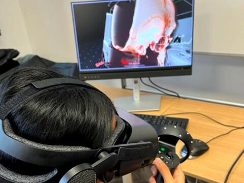
At Sorbonne University, the Faculty of Medicine's Simulation Department is preparing practical sessions in which 3rd-year medical students (DFGSM3) learn anatomy in three-dimensional (3D) situations, using virtual reality. These sessions are held in small groups of ten students over two semesters, with a virtual reality headset and a joystick held in the hand.
The simulation department has acquired three virtual reality workstations using AVATAR MEDICAL® technology. This technology uses medical images (CT scan, MRI, ultrasound, etc.) to produce a fluid, high-resolution 3D virtual image (also known as an Avatar) almost instantaneously. Students can thus immersively explore the structures of interest (such as bones, vessels, muscles, viscera, and skin).
Sorbonne University is the first French university to be equipped with this technology. Dr. Jebrane Bouaoud is the coordinator of virtual reality projects within Sorbonne University's simulation department, and teaches Maxillofacial Surgery at the Pitié-Salpêtrière Hospital, a public hospital in Paris. Students interviewed during a tutorial session on maxillofacial anatomy and traumatology say that "the 3D virtual reality visualization that we can easily manipulate ourselves in space (helps them) compared to a standard 3D view that we can have on a simple computer screen. We can enter structures that are usually more difficult to visualize with a scanner. We're used to working on paper-based courses and it's much harder to understand, except when we're on a training course and it's explained to us clearly what we're seeing. We're told that there's this fracture and we're obliged to believe the teachers without even having seen the fracture being discussed, whereas here we see it ourselves, with 3D virtual reality, and it's much easier to understand."
Dr Jebrane Bouaoud and Jérémy Leroy from the Simulation Department
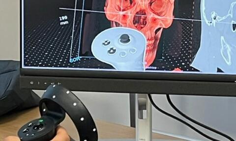
Close-up of the joystick and the 2D on-screen image


In practice, the teacher, Dr Jebrane Bouaoud, invites students to study cases of maxillofacial traumatology, such as a case of facial fracas (multiple facial fractures). During the tutorial session, students take on the role of a young maxillofacial surgery intern on call for the Île-de-France region. The clinical case is presented and the imagery (in this case cranial and facial CT scans) is analyzed by the students. The teaching objective is to provide a precise lesion diagnosis after analyzing the various fracture features. This hands-on immersion helps students, who have received classic theoretical courses at the start of the year, to gain a better understanding of maxillofacial traumatology. For example, as one student put it: "We already know the basic structures, and for example, just now there was an element of the course where we'd learned the basic idea, but seeing it was much more intuitive. It teaches us practical anatomy, with visualization of certain bone structures that are much more difficult to see on the standard scanner. It's a very good teaching tool, because it's sometimes hard to understand what you read in a book. What's more, there's a teaching team on hand to explain what we're seeing. We really enjoy the experience.
The students' anatomy learning curve also seems to be accelerated. Indeed, as Dr. Jebrane Bouaoud states, "senior doctors and interns have trained their brains for many years to try and visualize a 3D image in space from a standard scanner. For younger people, especially students who don't have this experience, the use of virtual reality tools such as Avatar Medical® facilitates this 3D reconstruction in space."
Do you have an idea for a virtual reality project? Don't hesitate to contact us.
Dr Jebrane Bouaoud
Virtual Reality Project Coordinator, Sorbonne University Simulation Department (DÉESSES)
jebrane.bouaoud@aphp.fr
AVATAR MEDICAL is an innovative spin-off from Institut Pasteur and Institut Curie, created in Paris in July 2020 by an experienced team of French and American co-founders.
