Seeing with Sound
Restoring sight to blind people thanks to a therapy that combines genetics and ultrasound is the hope of Serge Picaud's team, director of the Vision Institute*, and the Physics for Medicine Laboratory of the ESPCI** in partnership with the Institute of Molecular and Clinical Ophthalmology of Basel.

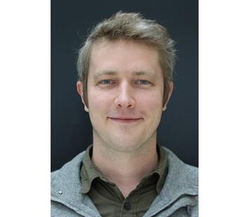
Gregory Gauvain is a associate professor at Sorbonne University and is attached to the Vision Institute. He participated in the development of this sonogenetic therapy, which consists of selectively activating certain genetically modified neurons remotely by ultrasound. He explains how this new brain-machine interface, tested in animals, represents a major innovation and a therapeutic hope for people suffering from optic nerve atrophy.
How is sonogenetics a major innovation for blind people, especially compared to the latest advances in optogenetics?
Gregory Gauvain: Contrary to optogenetic therapy, which focused on visual restoration at the level of the retina, sonogenetic therapy has the advantage of acting directly on the level of the visual cortex. Therefore, in theory, it could provide a solution to restore sight to patients who have lost the connection between their eyes and their visual cortex due to pathologies such as glaucoma, diabetic retinopathy, or hereditary or dietary optic neuropathies
How does it work?
G.C.: Sonogenetic therapy consists of genetically modifying certain neurons so that they can be remotely activated by ultrasound. Using gene therapy vectors, we introduce the genetic code of a membrane channel sensitive to certain ultrasound frequencies into the cells of a rodent's visual cortex. Only neurons in the treated area that express this channel can then be remotely activated by low-intensity ultrasound applied to the brain surface.
What did you demonstrate in the study published in Nature Nanotechnology?
G.C.: We have provided proof of concept of sonogenetic therapy on retinal neurons and visual cortex in animals. To test its effectiveness, we taught rodents undergoing water restriction an associative behavior between a flash of light and the arrival of a drop of water. Then, we replaced the visual stimulation (the light flash) by a sonogenetic stimulation in their visual cortex. We then observed the same reflex behavior, which suggests that the sonogenetic stimulation of their visual cortex did induce the light perception that caused the behavioral reflex.
If we convert the images of our environment into coded ultrasound waves to stimulate the visual cortex directly, at rates of several tens of frames per second, this therapy would be a hope for restoring sight to patients who have lost optic nerve function.
This is multidisciplinary work between the Vision Institute and the ESPCI. Can you tell us more about this collaboration?
G.C.: The collaboration with Michaël Tanter's team has enabled us to accelerate the project enormously. The team is specialized in waves in medicine and they are currently developing a functional ultrasound imaging system. The system will provide a visual representation of the brain activity induced by sonogenetic stimulation. This is a major advance for the development of sonogenetic therapy and more broadly for other neuroscientific innovations.
What steps are still to be taken before a clinical trial in humans can be considered?
G.C. : The development of a clinical trial still requires many steps to validate the efficacy and safety of this therapy. Among them, we will first have to prove that sonogenetic stimulation does indeed generate visual perception. For this, we will have to show—and we are already working on this—that the animal discriminates between two shapes with two different stimulations.
We will also have to verify on more complex animal models closer to humans, that we are able to stimulate the brain in depth and on specific areas, with a good spatial resolution.
At the same time, we will have to see how to create stimulation patterns according to each patient and be able to adapt the simulation in real time according to the measured activity. The functional ultrasound imaging system developed by Michaël Tanter's team will be precious.
Finally, for the clinical transfer, we will have to study, with industrial partners, how these strategies evolve over time and ensure the safety of the therapy.
*Sorbonne University/Inserm/CNRS
**ESPCI Paris/PSL University/Inserm/CNRS
